How Whipworms Tunnel Through the Gut

While often considered a neglected tropical disease, whipworm infections can occur all over the world—and have throughout history.
“We have archeological findings showing that these worms were infecting Vikings and Romans, and we actually found that the worms were inside Ötzi (the Iceman),” said Maria Duque-Correaan immunologist focusing on host-parasite interactions at the University of Cambridge.
Maria Duque-Correa uses gut organoids to study how whipworms interact with their hosts.
Jose Angel Dianes Santos
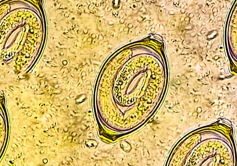
Different whipworm species infect people and animals when the latter consume food or water that’s contaminated with the parasite’s eggs. The worms hatch in the large intestine and then burrow into the epithelial cells of the gut where they develop into adult worms to produce more eggs that are shed in feces. The worms can stick around in the gut for years.
The human whipworm, called Trichuris trichuurahas evolved with people for millennia. Because of that, they trigger an immune response that—in an effort the get the worms out of the body—seems to counteract certain inflammatory conditions such as allergies and inflammatory bowel disease.1
“It’s not unheard of that people have tried to infect themselves with these parasites to try then to treat those conditions,” said Duque-Correa. “But because we don’t understand really what the worms are doing, and the worms actually do cause disease in 500 million people, it wouldn’t be wise just to say that we are going to go and infect everyone.”
There is no vaccine to prevent whipworm infections, and while there are antiparasitic drugs that can treat the parasite, they are not always completely effective. Duque-Correa and her team wanted to better understand how whipworms infect the gut to find new ways to prevent and treat these infections. To do this, they developed a gut organoid through which they watched whipworms invade gut epithelial cells in real time.2 Scientists can “use the new technologies to really understand how these worms live inside ourselves,” she said.
How To See a Tiny Worm in the Gut
To study whipworms in the lab, Duque-Correa uses the mouse-infective species, T. Mouse. While different whipworm species have specific animal hosts—from cats to dogs to primates—how the worms act in mice resembles their modus operandi in humans.
The challenge with watching T. Mouse inside a mouse is that the little whipworm larvae that hatch from the parasite eggs are only 150 micrometers long and hard to see against the many cells of the mouse gut. Unlike in studies of bacteria and unicellular parasites which researchers can genetically manipulate to express fluorescent markers, creating genetic knockouts or knock-ins is much more difficult in whipworms. With this limitation, Duque-Correa and her team thought that a gut organoid might be a better way to watch the early stages of how whipworms infect their host.
She started by developing a simple three-dimensional organoid consisting of gut epithelial cells. These organoids form with the apical side of the gut epithelial cells on the inside. Because whipworm eggs first encounter this side of the gut cells during an infection, Duque-Correa had to inject the worms into the center of the organoids.
“I did spend maybe a year of my life trying to microinject these parasites,” she sighed. One challenge she encountered was that while the worms are small, they are still orders of magnitude larger than bacteria, which meant that they regularly blocked the microinjection needles Duque-Correa was using to transfer the worms into the organoids. Also, the oxygen level inside the two-dimensional organoids was too low for the worms, so the ones that did get into the system died.
So, she shifted her focus to building a trans-well system to create a gut organoid.3 The benefit of this system was that she could access both the apical and basal side of the gut cells and could easily manipulate the system.
“We managed to recreate all the cellular populations we found in the intestine. When we put the worms (on the gut organoid), after a lot of trials, then they went inside cells and created the same tunnels that we see in the mice. So that was like our eureka moment,” she said. “That enabled us then to understand a little bit more how the worms live inside the cells.”
A larval-stage whipworm (light blue dots) infects intestinal epithelial cells (cell nuclei in dark blue) in a gut organoid. Tuft cells are shown in red, and the mucins in goblet cells are green. F-actin is stained white. Scale bars are 20 μm.
Maria Duque-Correa
The only problem was that they couldn’t live image parasites in the trans-wells. They had to fix the cells with the parasites in them, stain them, and then mount them on a microscope slide. But as luck would have it, Duque-Correa came across a paper from bioengineer Matthias Lütolf’s group at the Swiss Federal Technology Institute of Lausanne, where he and his team had grown organoids built on scaffolds that mimicked the gut structure. Her team initiated a collaboration with Moritz Hofera then graduate student in Lütolf’s lab, to see if they could build a gut organoid that would allow them to better model the infection process and watch the parasites in real time.
And after training Hofer to work with the parasites in the middle of the COVID-19 pandemic, the collaboration was a success.
“From that, we got the beautiful videos that actually are showing how the worms move inside the intestinal epithelia,” she said.2 Duque-Correa was most surprised to see how the gut cells responded as the worms tunneled through them.
“The cells don’t explode by having this massive worm to get through. They are still surrounding the worm, and in the very early days of infection, they actually try to stop the worm,” she said. Normally, cells use their lysosomes to encapsulate and kill intracellular pathogens that get into them. The researchers saw that lysosomes from the gut epithelial cells were attempting to fuse with the worm’s cuticle, which is a protective structure that encases the outside of nematodes. The worm withstood the attack, and the cells eventually died.
“It’s the host trying to respond to the worm, but then the worm winning the battle,” she said. “The organoids enable us to see this in a way that we cannot see in the host.”
More Complex Organoids, More Questions
Like most other parasites, whipworms have complex lifecycles. While Duque-Correa and her team saw the beginning steps of how the larval stage worms infect the gut epithelia, they now want to see if the worms can develop further in their gut organoid models.
“These worms are like little snakes,” she said. They molt four different times to become adult worms. “They are not only growing in size, but they are developing in(to) much more complex worms as they are molting,” she added.
To facilitate those experiments, she and her team are developing their organoid system to include more cell types commonly found in the gut including stroma cells and immune cells so that they can study the interactions between the parasites and these different cells.
She also hopes to use these organoids to develop new drugs to treat whipworm infections. The cure rate of albendazole, the most commonly used drug, is only about 15 to 20 percent, she said. “We don’t understand why this is the case because if you actually take a worm out and you put the drug on top of it, you will kill the parasite, but you will not kill the parasite if the parasite is inside the cell,” she explained. She wondered if perhaps the tunnel the worm forms inside gut epithelial cells could be protecting the worm from the antiparasitic drugs.
“There is a need to understand them much better to develop new drugs, but we can also learn so much from them,” Duque-Correa said. “It really illustrates how the organoids can bring new knowledge into the parasitology field.”


